“The research findings may improve the capability of medical imaging technologies by providing a pH-responsive contrast agent that is potentially more sensitive and selective than commercially available products.”
— Prof Liu Xiaogang, NUS Department of Chemistry
NUS chemists have developed a high performance contrast agent for magnetic resonance imaging (MRI) technology to improve cancer diagnosis.
Current commercially available contrast agents for MRI suffer from two main drawbacks; they are unable to bind specifically onto cancerous tumours and have a short circulation time within the body. The signal-to-noise ratio is often low and this results in MRI images which have poor spatial resolution. It remains challenging to develop biocompatible contrast agents which can target cancerous tumours (with high selectivity) and provide imaging signals with high signal-to-noise ratios so that higher quality images can be obtained for medical diagnostic purposes.
MRI technology is a non-invasive medical imaging technique for disease diagnosis and therapeutic monitoring. It uses chemical compounds known as contrast agents to improve the contrast between the internal body structures and surrounding tissues.
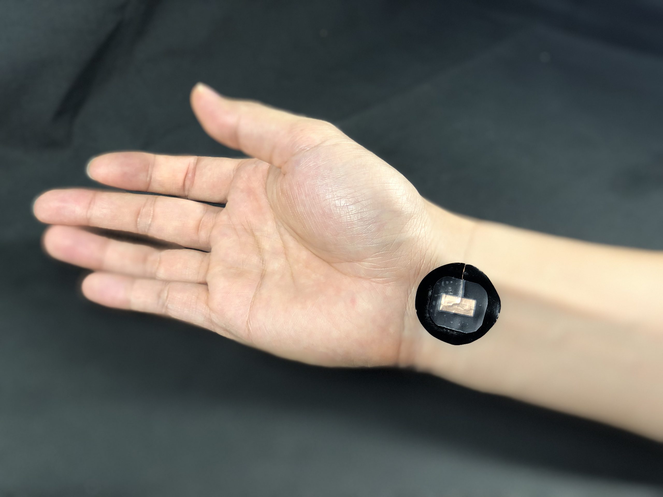
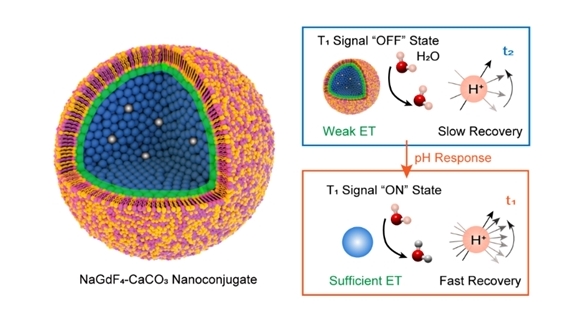
Prof LIU Xiaogang and this team from the Department of Chemistry developed the new class of nanoparticle-based contrast agents, known as MSNPs using self-assembled NaGdF4 and CaCO3 nanoparticles. Using a self-assembly process, the NaGdF4 and CaCO3 nanoparticles are agglomerated into spherical cores. Each of these cores is encapsulated by a layer of lab cultivated cell membrane. Under normal conditions in the body, the MSNP is not activated and gives an “off” signal. However, when the microenvironment is mildly acidic, which is the case for most cancer tumours, it becomes activated. Under this condition, the CaCO3 compound in the MSNPs reacts with water molecules present in the body to form carbon dioxide bubbles. This causes the disintegration of the MSNPs, releasing the NaGdF4 nanoparticles into the surrounding cancerous tissues. This results in an “on” signal. Based on experimental results using murine models, the MSNPs show a contrast enhancement of potentially more than 60 fold improvement in tumour visualisation when compared to Magnevist, a commercially available MRI contrast agent.
The research findings were published in Advanced Materials in 2019.
Yi Z; Luo Z; Barth ND; Meng X; Liu H; Bu W; All A; Vendrell M; Liu X*, “In vivo tumor visualization through MRI off‐on switching of NaGdF4-CaCO3 nanoconjugates” ADVANCED MATERIALS Volume: 31 Pages: 7 DOI: 10.1002/adma.201901851 Published: 2019.